Education
Three years of education are required to become a biomedical laboratory scientist in Norway. That will get you a bachelor’s degree, the title “Bioingeniør” and a valid HPR number (authorised health personnel number). There are seven different educational institutions in Norway; all with varying structures, but with the same national guidelines regarding learning outcomes. The biomedical laboratory scientist education consists of both theoretical as well as practical modules. The practical modules, on which the student can spend up to three months at a given department, take place at different hospitals all across Norway and include a mixture of several disciplines. During the third year the student has the opportunity to spend eight weeks in a specialist field, gaining even more in-depth knowledge. For the pathology module of the study, the student could come across the following disciplines: grossing, processing, embedding, sectioning and the final evaluation of slides. During the microtomy modules, the students get familiar with various microtomes.
Hanae Bourass has spent eight weeks at a pathology department for her final practical module. “It’s really great that you get to use the microtomes that are available on the market,” explains Hanae. “We were introduced to the old-fashioned sledging microtomes, the semi-automated microtomes and also the waterfall microtomes during the practical module.”
Work experience
“I completed my education in 2016. Up to now, I have had approximately two years of experience working as a biomedical laboratory scientist. I finished my training and practical modules at one hospital in the Oslo area, and have now started working at another hospital in the same area. What struck me most upon completing my education was the conservatism and scepticism regarding the usage of the different microtomes available in the pathology department.”
Different microtomes
“Working in a pathology department means working in an area with a long-standing tradition about how to use the equipment. It is also a field not at all dominated with huge technological improvements; for instance, like those we see in other areas of laboratory medicine. Where they were 10 years ago, is where pathology is today. Especially when it comes to equipment. Let’s take the different microtomes into perspective.”
Sledging microtomes
Hanae continues: “The key advantage of sledging microtomes is that they do not need a dedicated power supply. So if the electricity shorts out, you will still be able to do your sectioning. That was really appreciated 40 years ago, but today we have ‘emergency power’ which solves those challenges.
“The disadvantage is that it is time consuming to orientate the block. Can you really trust your eye? If the block is not orientated correctly, you risk losing valuable tissue. At the same time, as you section, your shoulders and neck are at risk of being injured as you manually pull the knife back and forth horizontally. We all want to take care of our own physical health and avoid as many work-related injuries as possible, which is also the reason why many departments have abandoned the sledging microtomes.”
Rotary microtomes
“Rotary microtomes are more or less the ‘gold standard’ in most Norwegian pathology departments today. The rotary microtome is semi-automatic, which reduces repetitive motion disorders, making it a more ergonomically attractive working instrument than the sledge microtome. A water bath is placed next to your microtome and, especially for non-trained or newly educated histotechnicians, the distance from the microtome to the water bath can seem never ending – and I assume we have all lost slides here. The slide can also get damaged if you grab it too hard with a pair of forceps. Then you’ll have to cut a new section, which is the reason why more and more scientists are using different kinds of brushes.”
Waterfall microtomes
“The main advantage of a waterfall microtome is the timesaving feature of having the water bath incorporated. Not only is it easy to pick up the sections, the risk of damaging the section during transfer is also reduced. Due to its integrated slide and incorporated water bath, we find that the waterfall microtome brings improved ergonomics because it eliminates the often-difficult transportation of the section from the knife blade to a separate water bath.
“The main challenge of a waterfall microtome is its transportation slide. This slide can get stuck and you will have to abort your job. It works all right if you haven’t started yet, but once a section moves down the slide and it stops working, it will take you a lot of time and energy to resolve the issue. Likewise, residual paraffin can become hidden between the blade and transport tape, which also needs to be removed for optimal speed of the waterfall over the slide. More seldom, we have experienced issues with the pumping mechanism, which will sometimes cause a water stop and make sectioning almost impossible. Still, you have to take into consideration that the more technical an instrument gets, the more your skills will have to change accordingly.”
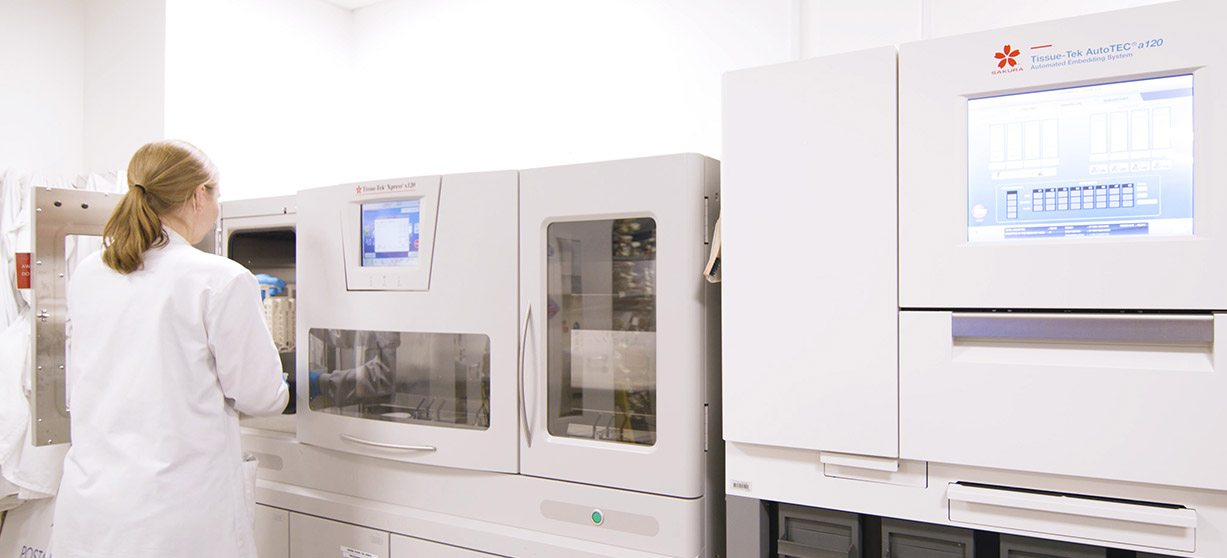
Tissue-Tek AutoSection®
The Tissue-Tek AutoSection is a fully automated microtome, where alignment of the block is done without any user interference. “Whether you are trimming or sectioning a new block or performing recuts from a previously sectioned block,” explains Hanae, “the AutoSection will do it all for you with just a push of a button. It’s something you have to get used to, spending your time watching and waiting, with your arms and shoulders in a downwards position – just relaxing. There is no need to focus, no ducking your head between your arms trying to adjust the block, no screws to be tightened or hand wheel to push. There is no stress... just an ergonomically attractive microtome.”
Hanae continues: “We use AutoSection primarily to trim blocks that have been embedded on the Tissue-Tek AutoTEC a120® through the Paraform® Sectionable Cassette System. The AutoSection is programmable, and we have pre-programmed it to remove the plastic inserts to make the blocks ready for sectioning. It saves time to have one person in the lab just trimming and making the entire production ready for sectioning. When sectioning blocks are embedded by the AutoTEC a120, all blocks are embedded with the same angle and in the same manner. Therefore, we spend less time adjusting the block-holder for subsequent blocks. Not only does this save time in the laboratory, it lends ergonomic help to the user’s shoulders, neck and back.
Main challenges in sectioning
You must always bear in mind that the important things to remember in sectioning are:
- Cut a good section without any holes or scratches
- Hard tissue and calcification impact the quality of the slide
- Manual embedding may play a role, and have an impact
There are several instruments on the market today, and all of them offer different features.
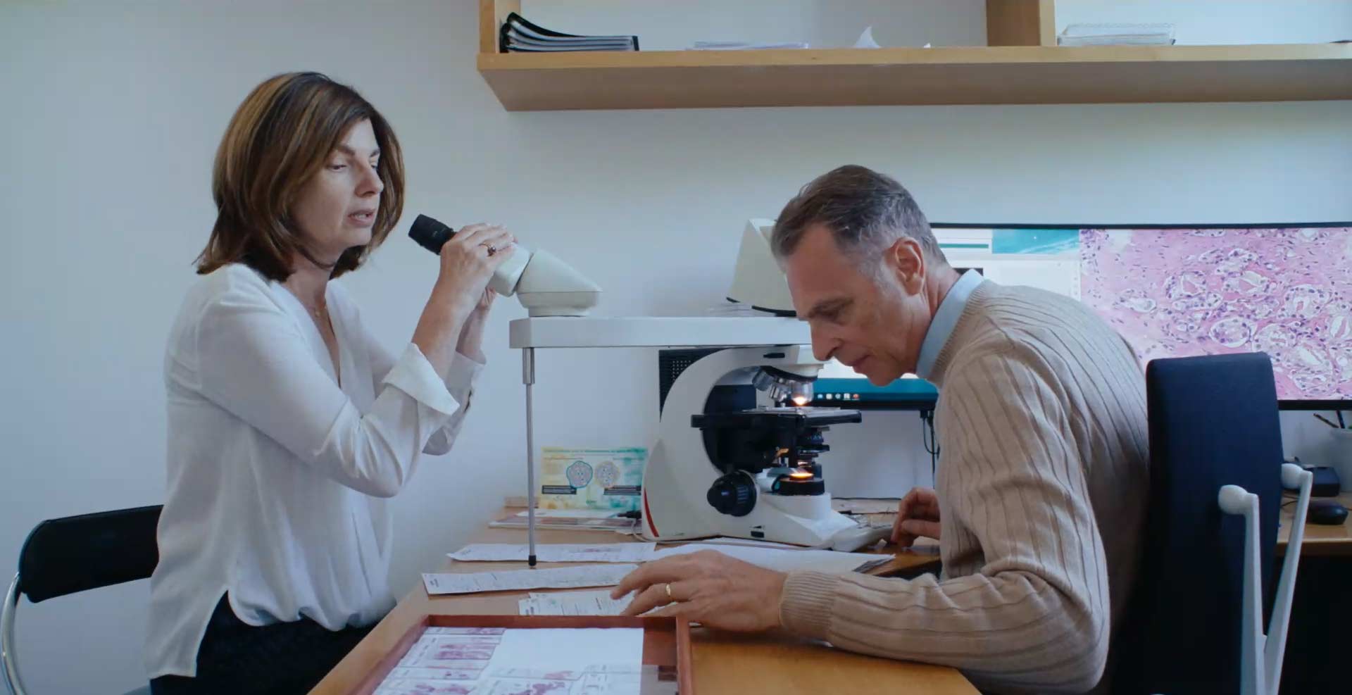
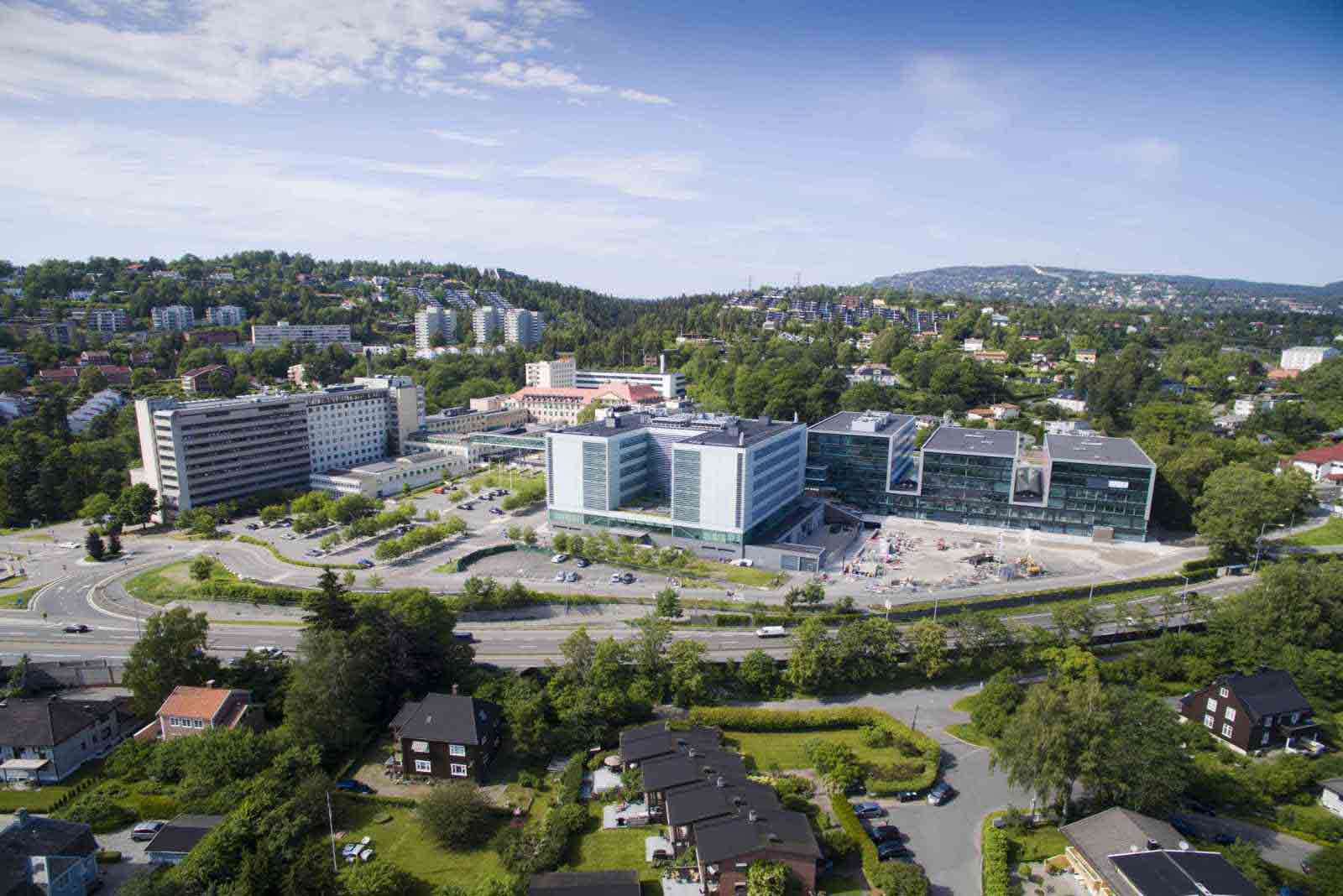
Department of Pathology, Oslo University Hospital
The Department of Pathology has broad competence and provides diagnoses on tissue and cell samples from inpatients and outpatients alike. Oslo University Hospital is a Comprehensive Cancer Center. The department operates out of the Radium Hospital, Rikshospitalet and Ullevål Hospital.
The department processes about 115,000 samples per year. These samples come from biopsies, surgical specimen, exfoliative cytology, aspirates and fine needle aspiration cytology. All samples undergo routine treatment, but we also have a wide range of special analysis such as electron microscopy, flow cytometry, immunohistochemistry, immunocytochemistry, molecular pathology and ploidy analysis. The Department of Pathology also includes extensive research and investigation activities. Our staff is subspecialized and the different locations have a somewhat different profile based on the type of specimens handled at each location. Unit of histology, Radium Hospital.
The unit of histology located at the Radium Hospital processes about 16,000 samples a year which includes 93,000 blocks and subsequently 110,000 H&E slides and about 10,000 special stains. The Norwegian Radium Hospital is Norway's leading cancer hospital and process gyneacological, urological, bone and soft tissue, breast and haematological samples for histology.